6 Patterns > Acute Lung Injury Pattern > Subpatterns > w/ Background Fibrosis
Acute Lung Injury w/ Background Fibrosis
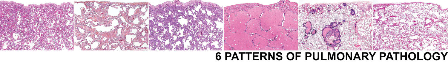
Surgical lung biopsy with evidence of both acute and chronic interstitial lung disease can be some of the most difficult to interpret. They are best approached by attempting to identify the dominant background fibrosis pattern (UIP, airway centered, or NSIP) and then to characterize the acute lung injury component (OP, fibrin, DAD). There are 4 main settings that you encounter this overlap pattern (in order of frequency)
CTD associated ILD undergoing a flare.
Acute exacerbation of UIP of IPF.
Chronic recurrent aspiration.
Subacute and chronic HP.
Flare of CTD Associated ILD
Chronic recurrent aspiration
Acute exacerbation of UIP of IPF
Subacute on chronic HP
Sample Signout
If no additional specific histologic features are identified, consider the following approach to signing the case out:
Acute on chronic interstitial lung disease (see comment).
Comment: The biopsy shows histologic evidence of both acute and chronic ILD. The predominant pattern of the acute component is _______ (select OP, DAD, fibrin, etc). The predominant pattern of the chronic compoennt is ______ (select UIP, NSIP, airway-centered, etc). No additional specific histologic features to indicate an etiology are identified. There is a broad differential diagnosis including acute exacerbation of UIP, flare of CTD, subacute on chronic HP, and acute on chronic aspiration. The diagnosis of ILD requires a multidisciplinary approach. Correlation with imaging studies and clinical history is suggested.