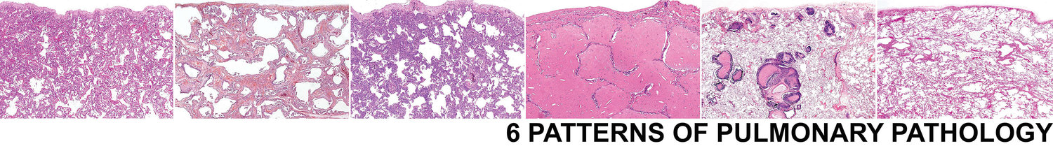
6 Patterns > Acute Lung Injury Pattern > Subpatterns > w/ Hayline Membranes > CTD Associated DAD
Connective Tissue Disease Associated DAD
…UNDER CONSTRUCTION…
Connective tissue disease (CTD) is a common cause of an acute lung injury pattern, including DAD. Histologic features in a case of DAD that should suggest CTD include the following:
Interstitial lymphoplasmacytic inflammation
Pleuritis
Lymphoid aggregates/follicles
Germinal centeres
Chronic bronchiolitis
SEE BELOW FOR SAMPLE SIGNOUT
If you are considering a diagnosis of CTD associated DAD, the biopsy should show the following features:
Hyaline membranes
Edema
Organization
Lymphoplasmacytic interstitial inflammation
Pleuritis
Lymphoid follicles
Interstitial Inflammation
Germinal Centers
Hyaline Membranes
Lymphoid Aggregates
Pleuritis
Chronic Bronchiolitis
Biopsies with the following features may not be infection associated DAD:
Interstitial giant cells or poorly formed granulomas (consider chronic HP)
Foamy macrophages and type II pneumocytes (consider drug reaction)
Foreign material (consider aspiration or intravenous)
Alveolar hemosiderin laden macrophages and capillaritis (consider alveolar hemorrhage syndrome)
Background fibrosis (consider acute on chronic ILD)
Necrosis (consider infection)
Viral cytopathic effect (consider infection)
Abundant neutrophils (consider infection)
Necrosis (consider infection)
Neutrophils (consider infection)
Foamy macrophages and type II pneumocytes (consider drug reaction)
Alveolar hemosiderin laden macrophages and capillaritis (consider alveolar hemorrhage syndrome)
Viral Cytopathic Effect (consider infection)
Interstitial giant cells or poorly formed granulomas (consider chronic HP)
Foreign material (consider aspiration or intravenous)
Background fibrosis (consider acute on chronic ILD)
Clinical Presentation
These patients often have a history of CTD and may have a chronic respiratory issue.
Flares of CTD may result in an acute worsening of symptoms
Although females are more often affected by CTD, males get CTD associated ILD more commonly
Often have elevated CRP and ESR and positive autoantibodies
Negative serology does not exclude the diagnosis as up to 20% of patients may not have autoantibodies at the time of diagnosis.
Radiology
Bilateral ground glass opacities
May have some evidence of chronicity in the form of reticulation
Sample Signout
If you are suspicious for an infectious etiology for the ALI biopsy, consider the following approach to signing the case out:
Acute and organizing diffuse alveolar damage with ______(insert necrosis, neutrophils, viral cytopathic effect, and/or granulomas) highly suspicious for an infectious etiology (see comment)
Comment: The biopsy shows an acute and organizing acute lung injury process. The presence of _______(insert necrosis, neutrophils, viral cytopathic effect, and/or granulomas) is suggestive of an infectious etiology. AFB and GMS (and IHC if viral cytopathic effect) stains have been ordered on multiple blocks. Regardless of weather they are positive or not, infection is still favored. Correlation with final culture studies and infectious serologic studies is suggested.
If the the biopsy shows DAD without any additional specific histologic features, consider the following approach:
Acute and organizing diffuse alveolar damage (see comment).
Comment: The biopsy shows acute and organizing diffuse alveolar damage. There is a broad differential diagnosis including infection, drug reaction, CTD, aspiration, and as an idiopathic entity. No additional specific histologic features to indicate an etiology are identified. AFB and GMS stains have been ordered on multiple blocks. If the patient is immunocompetent, the absence of necrosis, neutrophils, viral cytopathic effect, and granulomas makes infection less likely, although it cannot be excluded. The diagnosis of ILD requires a multidisciplinary approach. Correlation with microbiology studies, imaging studies and clinical history is suggested.