6 Patterns > Fibrosis Pattern > Additional Features > w/ Honeycomb Only
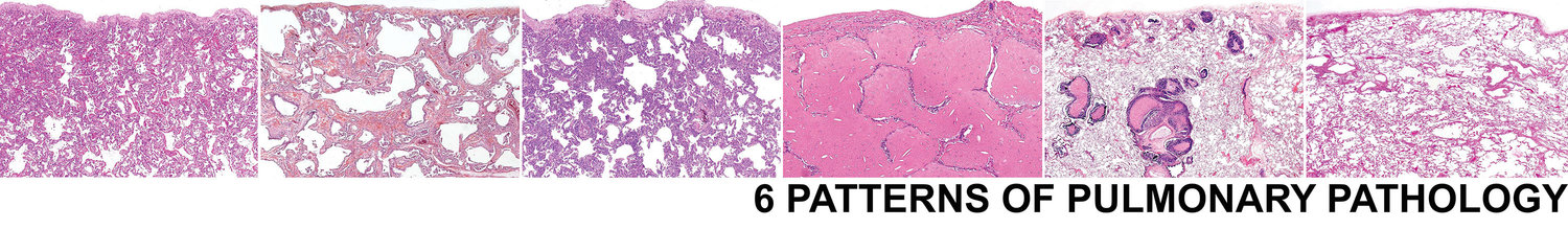
Fibrosis w/ Honeycomb Only
Honeycomb is end-stage pulmonary fibrosis and may be seen in ANY advanced fibrosing intersitial lung disease. It is similar to describing a liver biopsy as cirrhosis. This tells your clinician that there is end-stage fibrosis, but does not tell them anything about the etiology of the fibrosis.
There are 4 histologic features of microscopic honeycomb remodeling:
- Advanced scarring
- Dilated cystic spaces embedded in the scar
- Lined by ciliated epitheluim
- Filled with mucinous material

Advnaced scarring

Dilated cystic spaces in scar

Cysts filled with mucinous material

Cysts lined by respiratory epithelium
Knowledge of the CT imaging studies is essential in the setting of honeycomb only.
Diffuse Disease on CT
With diffuse disease on CT, and a biopsy with honeycomb, you can be confident in a diagnosis of chronic ILD. CT distribution can help point to an etiology, as can the following features:
- Honeycomb without any other features > UIP of IPF
- Giant cells and granulomas > Chronic HP and sarcoid
- Marked Lymphoid hyperplasia (you may allow some degree of lymphoid hyperplasia in any advanced fibrosing process) and pleuritis > CTD
- Abundant elastosis > PPFE
Localized Disease on CT
If localized on CT, this is NOT a chronic ILD and consider the following:
- Middle lobe syndrome
- Recurrent infection
- Aspiration
- IgG4 disease
- Congenital anomaly
Sample Signout
If no imaging studies are available, consider the following approach to signing the case out:
End-stage lung with microscopic honeycomb remodeling (see comment).
Comment: The biopsy consists of advanced fibrosis with microscopic honeycomb remodeling. If the imaging studies show diffuse interstitial lung disease, this likely represents an advanced fibrotic ILD. The 2011 ATS/ERS classification system suggests assigning this biopsy as a "Probable" UIP-pattern. Many different ILDs could be responsible for the end-stage fibrosis seen in this case.
If the CT shows a localized lesion, this could be related to middle lobe syndrome, recurrent infection, aspiration, IgG4 disease, or a congenital anomaly. Correlation with imaging studies and clinical history is suggested.