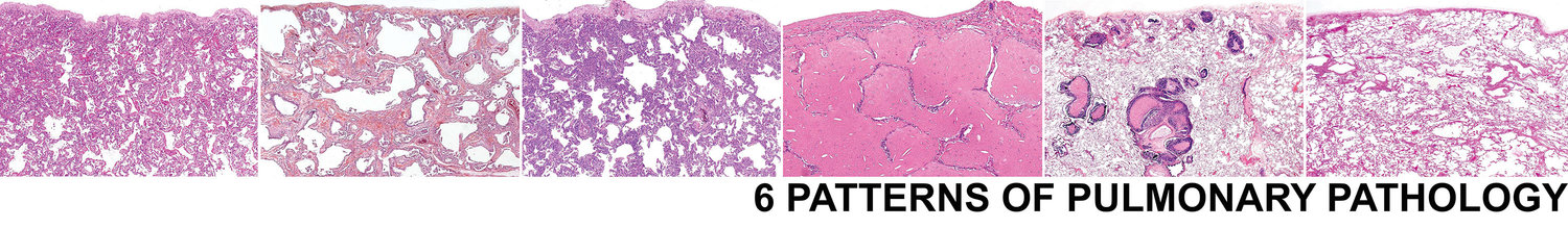
6 Patterns > Fibrosis Pattern > Additional Features > w/ Diffuse Alveolar Wall Fibrosis > Smoking Related Interstitial Fibrosis
Smoking Related Interstitial Fibrosis
Smoking related interstitial fibrosis (SRIF) is one of the best mimics of fibrotic nonspecific interstitial pneumonia. In fact, many classic text book images of fibrotic NSIP actually are cases of SRIF.
SRIF may be encountered incidentally (in lobectomies for tumor) or may be a part of a diffuse interstitial lung disease related to smoking. The term smoking related interstitial lung disease (SR-ILD) encompasses a variety of histologic entities including respiratory bronchiolitis (RB), desquamative interstitial pneumonia (DIP), pulmonary Langerhan cell histiocytosis, emphysema (PLCH), and SRIF. This term should only be used in the setting of respiratory compromise clinically and with radiographic evidence of ILD.
SEE BELOW FOR SAMPLE SIGNOUT
If you are considering a diagnosis of SRIF, the biopsy should show the following features:
Diffuse expansion of the intersitium
Densely collagenized fibrosis
Simplification of the alveolar wall architecture in the background (emphysema)
RB is commonly encountered
Diffuse expansion of the interstitium
Simplification of the alveolar wall architecture in the background (emphysema)
Densely collagenized fibrosis
RB is commonly encountered
SRIF should be questioned in the following settings:
Lymphoplasmacytic interstitial infiltrate (consider CTD)
Loose elastotic interstitial expansion (consider CTD)
Lack of other smoking related changes (emphysema, RB)
Abundant dust and inhalational debris (consider pneumoconiosis)

Lymphoplasmacytic interstitial infiltrate (consider CTD)

Lack of other smoking related changes (emphysema, RB)

Loose elastotic interstitial expansion (consider CTD)
Abundant dust and inhalational debris (consider pneumoconiosis)
Sample Signout
The signout of cases with SRIF depends on the reason you are looking at the lung tissue under the microscope. If you encounter SRIF in the setting of a lobectomy for tumor, simply include SRIF as an incidental finding (unless the patient also has clinical and radiologic evidence of an ILD).
If you have a biopsy for ILD and the predominant finding is SRIF, consider the following approach to signing the case out:
Extensive smoking related interstitial fibrosis (see comment)
Comment: The predominant histologic abnormality in this case is SRIF. SRIF may be an incidental finding or may be a component of smoking related interstitial lung disease (SR-ILD). Although there are minimal other changes of smoking on this biopsy, in the correct clinical and radiologic setting the biopsy would be compatible with SR-ILD. Correlation with the clinical history and radiographic imaging studies should help confirm the diagnosis.
If there is extensive SRIF in the setting of other smoking related pathology (RB, PLCH, DIP, emphysema), consider the following approach:
Advanced smoking related lung pathology. In the correct clinical setting the changes are diagnostic of smoking related interstitial lung disease (see comment)
Comment: SR-ILD is an encompassing term to include the spectrum of interstitial lung disease seen in the setting of defensive tobacco smoking exposure to include RB, DIP, PLCH, SRIF, and emphysema. This case shows __________. Obviously, smoking cessation is of the utmost importance.